Psychology 1
PREVIOUS: Introduction to Psychology
Neurons
Everything psychological is biological.
Your brain releases chemicals when you’re scared or happy or whatever. All of your sensations, feelings, mind are all tied to your brain and the neurons. Like organisms, these sort of have a hierarchical structure:
- Cell :: Neurons
- Tissue :: Nerves, Ganglia, Nuclei
- Organ :: Brain, spinal Cord, Brainstem
- System :: Nervous System (Central, Peripheral)
- Organism :: Person
Neurons: also known as nerve cells, are the building blocks that comprise our nervous systems.
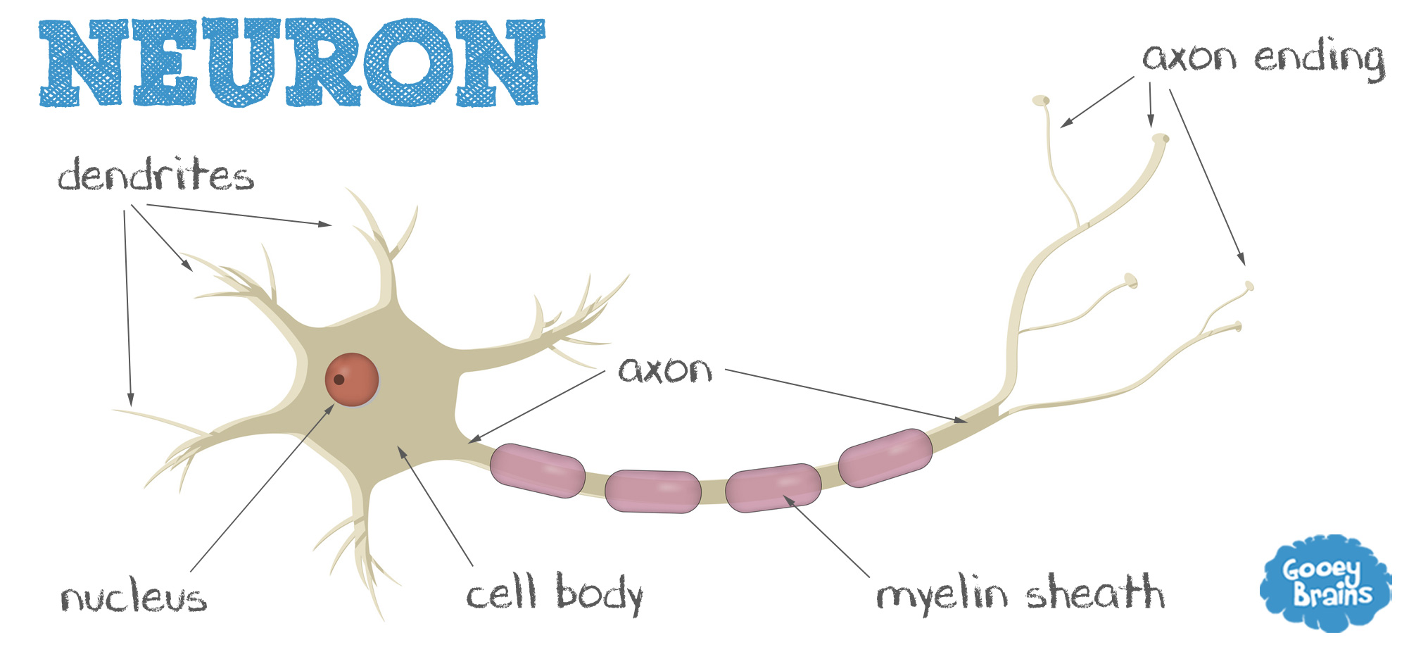
There are three types of neurons:
- Afferent: which control senses
- Efferent: which control our “motorary” movement
- Interneurons: Communication through the central nervous system
In robotics terms, the afferent would be your sensors, the efferent would be the actuators, and the interneurons are the control software.
Dendrites: “tree”, branching structures that receive inputs from other neurons. They’re the listeners that relay information to the cell body.
Axon: The talker. It releases electrical inputs for other dendrites to receive. What a great ROS publish/subscribe system.
Myelin Sheath: Depending on the neuron, some axons may be protected by the myelin sheath. This also speeds up transmission time of messages.
Neurons transmit signals when stimulated by sensory input or when they are triggered by neighboring neurons. The dendrites pick up signals and activate the action potential of the neuron, that shoots the charge down the axon to its terminals toward neighboring neurons.
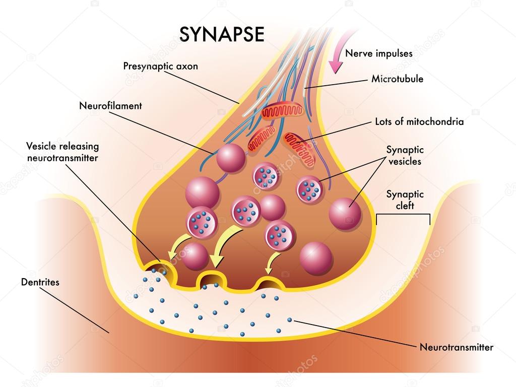
The sending neuron’s terminal axons and the receiving neuron’s dendrites are “connected” at the synapse. They don’t quite touch, but the millionth of an inch between them is called the synaptic cleft. The messengers that jump the gap are called neurotransmitters. But once the message is relayed, they go back to the original axons in a process called reuptake.
All of these control our emotions, thoughts, feelings! We have hundreds of different types of neurotransmitters, but the split into two different branches: excitatory or inhibitory. Here’s one.
Endorphins: natural, opiatelike neurotransmitters linked to pain control and pleasure.
A lot of famous “mental illnesses” are linked to these ideas. Alzheimers, schizophrenia, seizures, depression, are all linked to the different lack of or excessive neurotransmitters in our brains.
And drugs! Stimulants can increase energy, alertness, and activity by preventing the process of reuptake (leaving excitement longer), or stimulating synapses, etc.
Note that axons can not change the strength or velocity of its action potentials. This is known as the all-or-none law.
The Nervous System
This is the structure of our Nervous System.

This is where the afferent/efferent neurons that were previously described come into play. The Afferent neurons send sensory information up to the brain, who processes what to do, and sends signals down the spinal cord on which motors to use. Since messages are relayed through the spinal cord/somatic system, damage to the spinal cord will cause loss of function and sensation–paraplegia.
The General Stress Adaptation Syndrome describes how stress plays into our life. When we first experience stress, our sympathetic (NS->PNS->Sympathetic) part of our NS activates, taking on the “fight or flight” mode. But as stress continues, we experience decreased emotionality, because our parasympathetic NS kicks in. Continued stress will result to exhaustion and potential death, as our body depletes its resources.
The Brain
Most of our brain research today are based on the study of lesions. These are injuries to the brain that impair certain functions, so we can then take scans of the brain and see which part is damaged and sort of map out which part of the brain controls which aspects of our lives. We can also create temporary lesions by cooling or shocking the brain with electrical current.
For example, Broca found that people with impaired speech have damage in one specific part of the brain, now known as Broca’s Area.
We can also study psychophysiology, which measures the nervous system–like lie detectors from the movies. Technology also now supports brain imaging (hi vivan) and reconstructs a 3D model of the brain. Through these, we can see which parts of the brain on certain mental tasks–happy thoughts, irritating thoughts, etc.
People have done research on trying to recreate what a person is viewing based on their brain scans.
Doctrine of Modularity:
The brain is highly specialized. Jerry Fodor supported this idea. Different mental functions are served by different mental organs that are dedicated to those specific purposes. These mental organs are associated with certain tasks. For example, speech is related to Broca’s Area. There are different processes for visual stiumlation or language processes–this is one of the newest tasks for neurosciences today. Informational encapsulation–the operation of a module is uninfluenced by any other module. Everything is independent and separate from each other. They are HARDWIRED. This is YOUR JOB. They are INATE. They are NOT acquired by experience or learning. There is also a suggestion that there are specific times for development–language processing is learned a specific time in your life. There is a fixed neural architecture: it is associated with a particular structure or system in the brain that is dedicated to performing the function of that module.
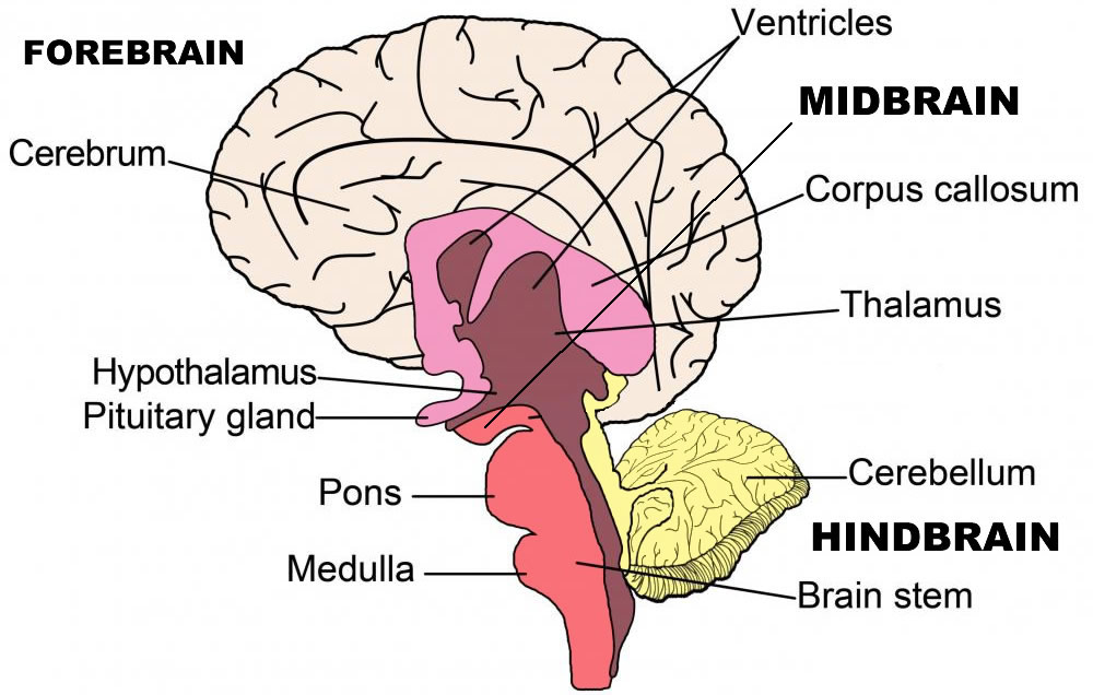
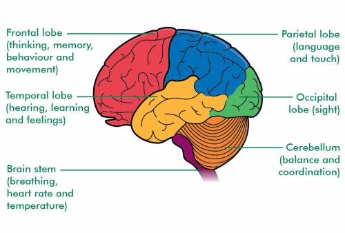
Parts of the Brain
- Hindbrain: controls coordination and is made up of
- Medulla: vegetative functions (heartrate, breathing)
- Pons: cortical arousal (cycle of waking and sleeping)
- Cerebellum: sensation and action (balance, coordination)
- Midbrain: consciousness, damage can lead to coma or vegetation.
- Reticular Formation: cortical arousal (similar to pons)
- Forebrain:
- Cerebal Cortex: what we think of ‘brain’
- Center of the Brain: (limbic system, basal ganglia, diencephalon)
- Hippocampus: Memory!
H.M. Had a part of his hippocampus removed because of his seizures. They stopped, but he was no longer able to retrieve memories. He would meet people, work on the same puzzles, and have no recollection of anything he’s done already.
- Amygdala: Emotions!
Patient S. had a disease that had calcification of her amygdala–it solidified so that it didn’t function as a structure. She had huge deficits in emotional responses–fear, inability to recognize facial expressions, disgust, etc.
- Hypothalamus: biological motives (hunger, thirst, parenting)
Lesions in parts of the hypothalamus disrupted eating behavior–rats that over-ate to obesity or starved themselves to death.
Triune Brain
MacLean’s Triune Brain is another way to organize the brain:
- The New Brain (neocortex)
- The Old Brain (limbic System, amygdala, hypothalamus, hippocampus)
- R-Complex (brain stem, cerebellum)
The Cerebal Cortex
We organize our Cerebal Cortex using Brodmann’s numbering scheme:
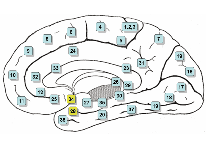
For example, the primary motor cortex is comprised of the precentral gyrus and premotor cortex, which correspond to Brodmann Areas 4, 6, and 8. The primary somatosensory areas (touch, heat, and cold) correspond to Brodmann’s area 1, 2, and 3.
We know this because we can analyze strokes. Lack of blood to a certain part of the brain can cause us to lose our primary motor cortex.
Aphasia: Problems with Speech
- Broca’s Aphasia: “Expressive”
- Slow, labored, in articulate speech (not fluent)
- Comprehensible
- No problems understanding speech or reading, but might have difficulties talking or writing.
- Wernicke’s Aphasia: “Receptive”
- They are fluent and sound Informational
- Words aren’t chosen properly to come across the correct meaning
- Might come out completely meaningless
- Problems understanding speech or writing.
Fusiform area for Facial Recognition
Brodmann’s Area 37. The people can recognize faces fine describe them, but can not identify people familiar to them–even themselves. They have damage in the Fusiform gyrus.
Hemisphereic Specialization, Recovery of Function, Plasticity
Our brain has two hemispheres: left and right. They each control the opposite side of the body–this idea is known as the contralateral projection. The right side of the brain controls the left side of your body and vice versa. But some parts are specialized also. Speech and language processing is clearly identifiable in the left hemisphere. So when aphasia occurs, it is because of damage in the left hemisphere. The left hemisphere is a little bit bigger, so we tend to prefer our right side, but there really is no difference.
Picture of a horse in the left visual field. This goes into the right hemisphere, but was not transferrable to the left hemisphere. The patient seeing the picture was able to draw the picture, but couldn’t say what it was.
Left hemisphere: language, sequential analysis, mathematical computation, fine motor control.
Right Hemisphere: Simple left-hemisphere functions, spatial analysis, pattern recognition.
How is knowledge represented in the brain?
- Localist View
- Individual pieces of information are stored in single neurons that are located near each other in the brain
- If you destroy that cluster of neurons, then you would obliterate the knowledge as well.
- Distributed View
- Every bit of knowledge is distributed throughout the cerebal cortex.
- Large number of neurons throughout a lot of space.
Recovery of function
Peripheral nervous system tissue can be recovered–if you cut off your finger and reattach it, it’ll still work. However, CNS tissue does NOT. However, there have been some cases where people with aphasia were able to recover. We consider those cases as “incomplete damage,” or that there might be other parts of the brain that can fill in on those broken parts of the brain. The idea of plasticity is that one part of the brain can take on the missing functions–our brain isn’t rigid and can reorganize itself. Or maybe CNS damage ISN’T permanent.